New Research Highlights Imaging Differences in Severe Forms of MS

New Research Highlights Imaging Differences in Severe Forms of MS Image

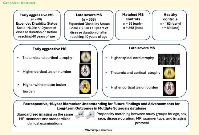
A new study published in Brain Communications by researchers at the University at Buffalo and the Buffalo Neuroimaging Analysis Center identifies distinct neuroimaging patterns in early aggressive and late severe multiple sclerosis (MS). Using 16 years of real-world MRI data and advanced AI techniques, the study found that early aggressive MS is linked to cortical and thalamic damage, while late severe MS shows more extensive spinal cord and grey matter atrophy.

These findings support more personalized monitoring and treatment approaches for patients with severe MS.

Full article: https://doi.org/10.1093/braincomms/fcaf25

Security Notice

The Buffalo Neuroimaging Analysis Center (BNAC) recently identified a cybersecurity incident involving unauthorized access to a portion of our systems. Upon discovery, we immediately took steps to secure the affected environment, engaged internal and external experts, and initiated a comprehensive investigation. At this time, the review process is ongoing. While a subset of data files may have been accessed, we are continuing to evaluate the scope and nature of the information involved. BNAC is working closely with appropriate legal, compliance, and regulatory authorities and will provide additional updates as more information becomes available. Individuals who may be directly impacted will be notified in accordance with applicable laws and requirements. We take the security of data entrusted to us seriously and are implementing additional safeguards to further strengthen our systems and processes. Updates will be posted to this notice as they become available.

For questions, please contact [email protected]


Interested in working
or training with us?

Schedule a meeting with our team.

Contact Us

Top