BNAC at ACTRIMS Forum 2025

BNAC at ACTRIMS Forum 2025
West Palm Beach, FL | February 27 – March 1, 2025
The Buffalo Neuroimaging Analysis Center (BNAC) demonstrated a strong presence at the 2025 Americas Committee for Treatment and Research in Multiple Sclerosis (ACTRIMS) Forum, sharing innovative work spanning the spectrum of multiple sclerosis (MS) research. The team delivered one platform presentation and 13 posters, engaging attendees with insights on neuroimaging phenotyping, glymphatic system dysfunction, proteomics, and connectome vulnerability. A central theme of the presentations was the application of advanced imaging techniques to characterize aggressive MS and track long-term disability progression. From paramagnetic rim lesions to novel metrics of brain clearance pathways, the BNAC team continues to bridge imaging biomarkers with clinical outcomes.
|
|
|
“This work fills a critical gap in MS research and high-lights the importance of addressing cortical pathology.”
|
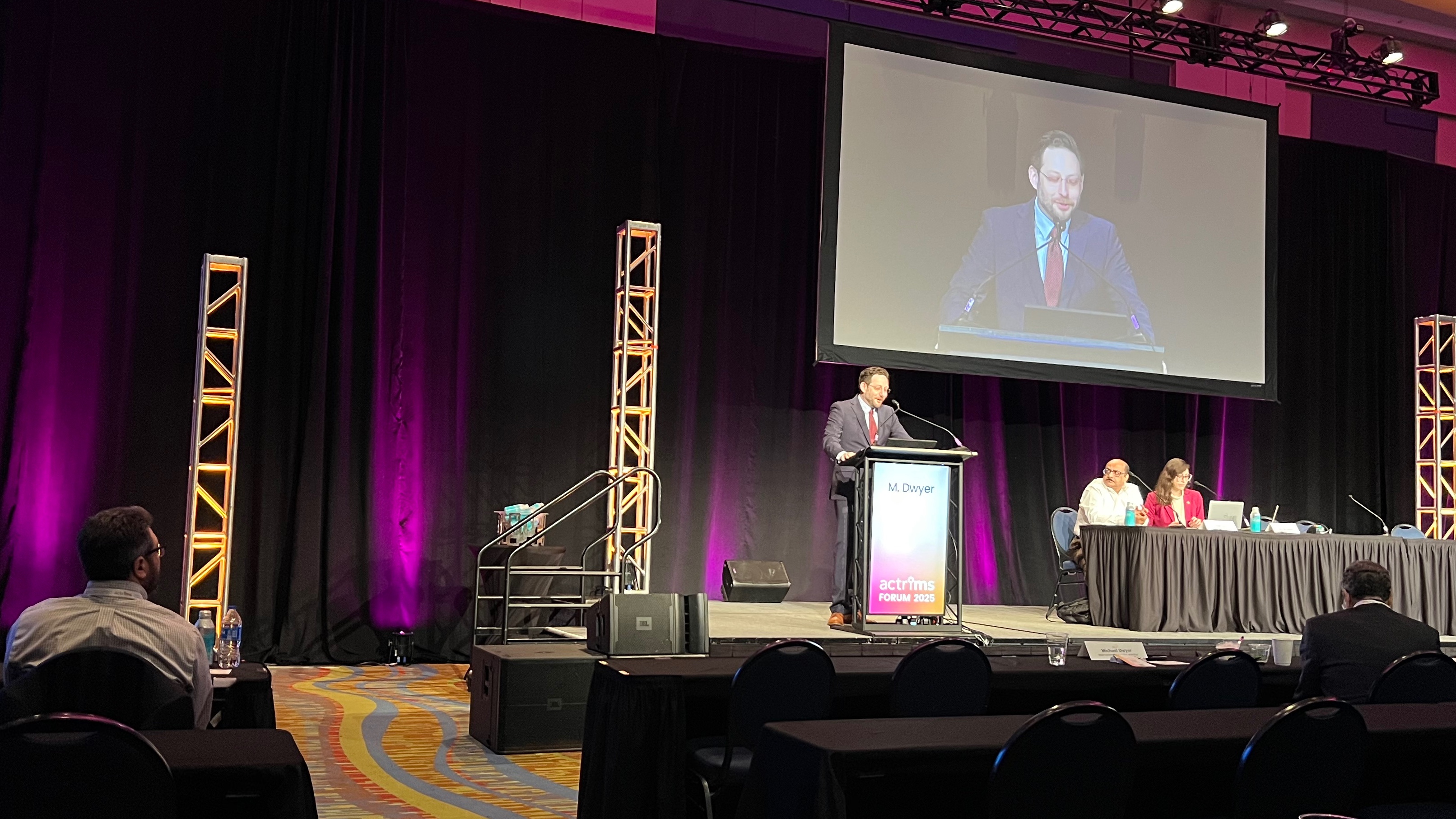
In one of the main platform sessions on Friday February 28, BNAC presented a landmark secondary analysis of the ORATORIO trial using advanced post-processing techniques to detect cortical lesions (CL) on legacy MRI. The study showed that treatment with ocrelizumab reduced new or enlarging cortical lesion activity by 74.8% and volume accumulation by over 86% compared to placebo over 120 weeks in patients with primary progressive MS (PPMS). This is the first large-scale, multi-center trial to demonstrate a therapeutic effect on cortical gray matter lesions—an area long considered difficult to study but highly relevant to disease progression. Dr. Michael Dwyer added, “ACTRIMS is always a great opportunity to share, learn, and connect with colleagues who are pushing the field forward.” BNAC’s collaborative energy was evident throughout the conference, as faculty, postdocs, and students stood side by side to present and discuss. The quality of research and diversity of methods presented captured BNAC’s commitment to excellence and innovation in neuroimaging science.
|
|
|
Ashley Tranquille shared new insights on PRLs and glymphatic function and presented findings from a 10-year longitudinal study on MS disability and glymphatic impairment. |
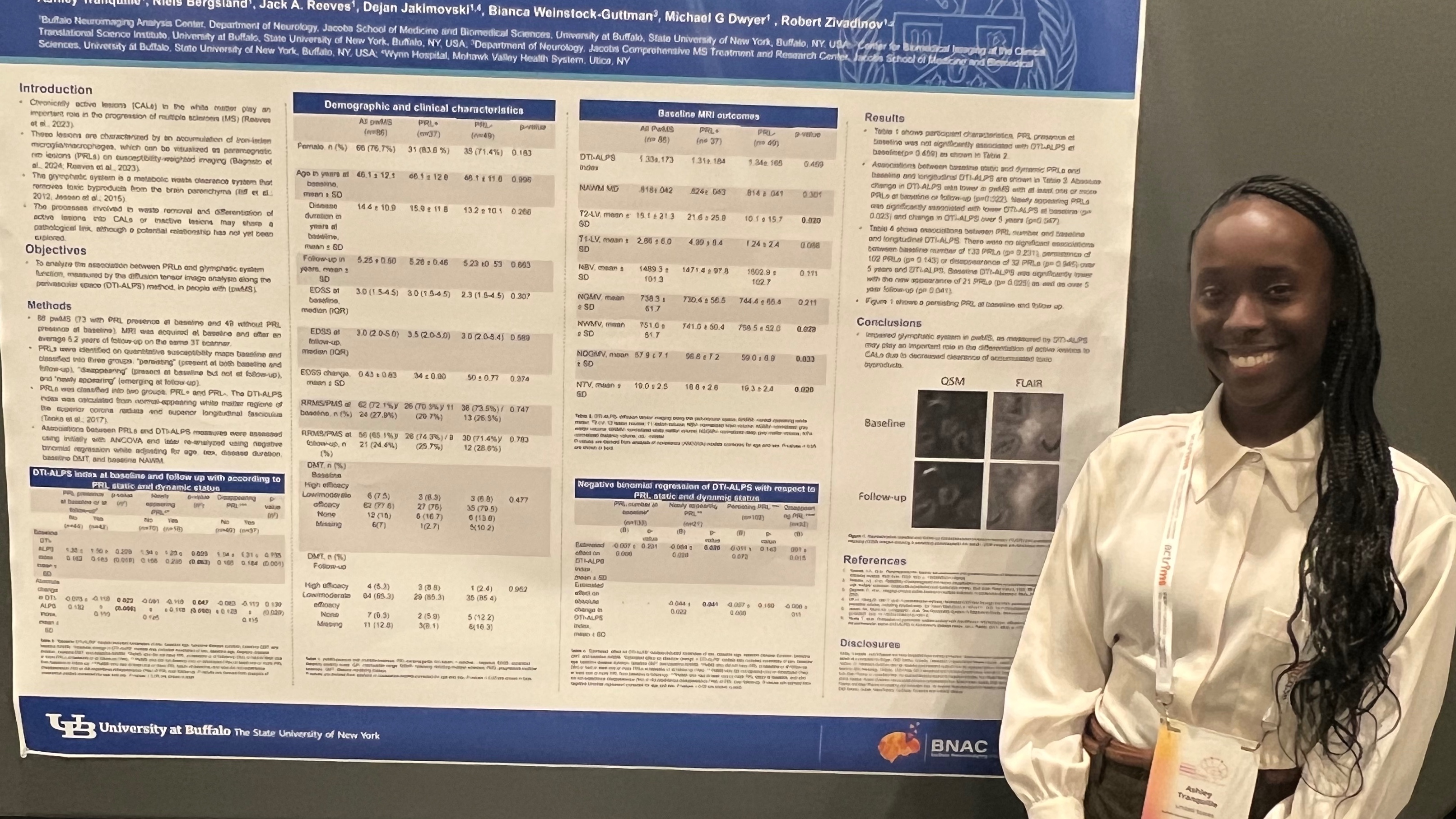
PhD student, Ashley Tranquille, presented compelling work on the glymphatic system’s role in MS, with a focus on how impaired waste clearance may contribute to long-term neurodegeneration. Her decade-long follow-up study revealed that reduced glymphatic function is significantly associated with accelerated disability progression, in progressive MS phenotypes. In a second presentation, she explored the presence of paramagnetic rim lesions (PRLs)—chronic active lesions visible on MRI—and demonstrated their strong link to glymphatic dysfunction. These findings underscore the emerging view that impaired clearance pathways in the brain play a central role in sustaining inflammation and injury in MS, beyond traditional white matter lesions.
|
|
|
Alexander Bartnik discussed imaging-derived connectomic vulnerability in MS. |
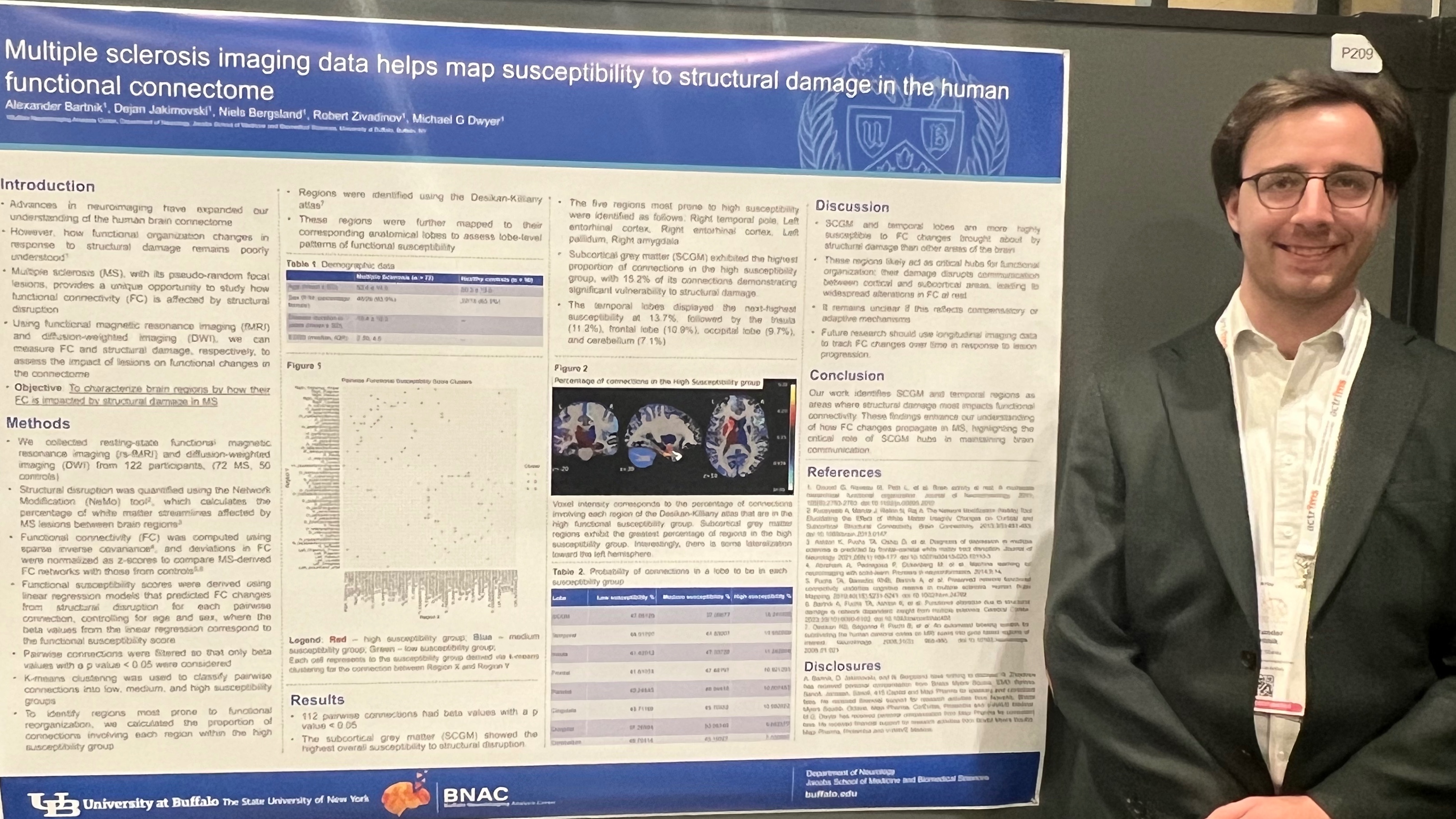
Dr. Alexander Bartnik, a post-doctoral fellow in BNAC, introduced a novel connectomic framework to explore how structural damage from MS lesions affects the brain’s functional network. Using advanced imaging techniques, his analysis identified highly connected “hub” regions that may be more vulnerable to lesion-related disruption. By mapping lesion location onto these hubs, Dr. Bartnik’s work offers a more precise way to understand which areas of the brain are at greater risk for functional decline. “This is a step toward personalized prediction of damage impact,” said Bartnik, highlighting the potential for this approach to inform individualized risk profiling and therapeutic strategies in MS.
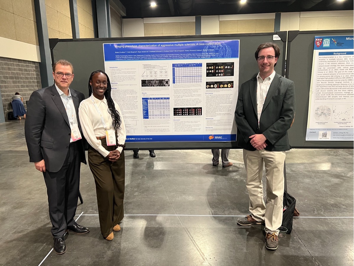
BNAC’s researchers analyzed early aggressive and late severe MS using data from the BUFFALO-MS database. In a cohort of over 900 participants, they found that people with aggressive/severe MS had significantly worse MRI outcomes than matched controls. Cortical lesion burden was the main driver of disability in early aggressive MS, while spinal cord atrophy, brain atrophy, and deep gray matter loss contributed to disability in late severe MS. The findings emphasize differing imaging phenotypes and drivers of disability across aggressive/severe MS trajectories.
|
|
Drs. Zivadinov, Tranquille, and Bartnik presented work on aggressive MS imaging phenotypes.
|
The future of BNAC’s research is bright. The entire team continues to advance the use of cutting-edge techniques to better understand MS, with the ultimate aim of helping to understand how to improve the lives of those affected by the disease.
###
Buffalo Neuroimaging Analysis Center | 77 Goodell Street, UB Downtown Getaway Building, Buffalo, NY 14203