Research Summary
prepared for the MS community by
Buffalo Neuroimaging Analysis Center
Advisory Council
Neuroimaging Correlates of Patient Reported Outcomes in Multiple Sclerosis
Dejan Jakimovski, Taylor R Wicks, Niels Bergsland, Michael G Dwyer, Bianca Weinstock-Guttman, Robert Zivadinov. Published in Degenerative Neurological and Neuromuscular Disease by Dove Press in 2023
MAJOR FINDINGS
Deep gray matter volume correlated best with MS disability v. lesion-load.
In surveys completed by participants it was shown that the volume of the thalamus was significantly influenced negatively by the level of reported depression
Patient-reported responses to questions about their symptoms on the simple questionnaire showed a surprisingly close correlation between deep gray matter volume, current disability, and likelihood for future disease progression.
Currently, MS Disability is assessed by two standards:
Physical performance testing
Clinicians traditionally measure MS disability using assessments that focus on physical, especially walking disability. The most common of these assessments has been the Expanded Disability Severity Scale (EDSS). However, EDSS scoring comes up short because of its narrow focus. For example, two people with MS may have identical EDSS scores, but report vastly different levels of fatigue, chronic pain, cognitive decline, bowel and bladder issues, and/or depression.
MRI analysis
MRI imaging for multiple sclerosis looks mostly at the number and intensity of lesions in the brain's white matter. This is referred to as white matter lesion load. This metric falls short because the experience of people with MS does not correlate with what is observed in clinic and MRI findings. For example, someone with a high white matter lesion load may have no clinical symptoms, while someone else with low white matter lesion loads may be significantly disabled.
The paper describes two better standards for assessing and predicting MS Disability
Deep gray matter analysis
Newer MRI analyses, such as measurement of the deep gray matter volume, have recently been suggested as the best radiologic markers of MS disability. As its principal finding, this paper concludes patient-reported outcomes correlate well with deep gray matter volume.
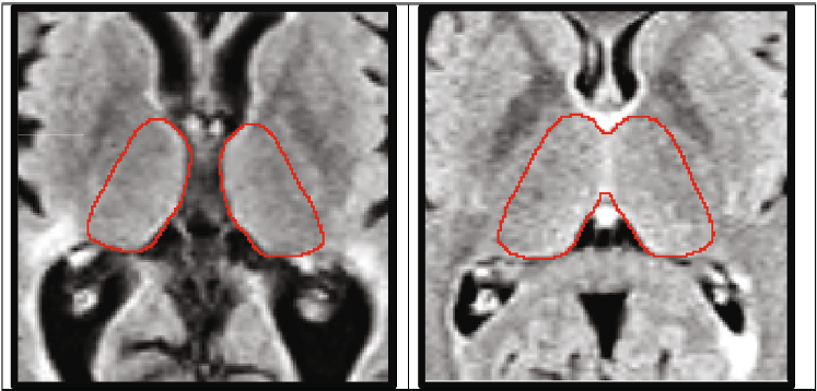
Above are examples of very high atrophy (left panel) and low atrophy (right panel).
Patient reported outcomes
This study used a patient-reported outcome questionnaire that compiled measurements that line up with what people with MS actually experience. This tool, currently used mostly for research, could also be used by clinicians as an additional way to track MS progression. Several papers published by the Buffalo Neuroimaging group over the past two years have shown that patient-reported outcomes are actually better at assessing patient condition and predicting future progression than more widely accepted metrics such as EDSS. Further research will be needed to understand how depression, a symptom reported by a significant number of people with MS, might affect disease progression.
Prepared by Advisory Council Members, Carol Schumacher, Mitchell Sturgeon, Tracie Jacquemin, Patricia Picco, Marc Stecker, and Craig Walters.
Click below for Advisory Council interview with Dejan Jakimovski
A Better Way to Measure Progressive MS

Print a copy of the research paper to share with your clinician
Print a copy of this Advisory Council Research Summary
Support MS Research at Buffalo Neuroimaging
Buffalo Neuroimaging Analysis Center Home Page
The Buffalo Neuroimaging Analysis Center is part of the Department of Neurology Jacobs School of Medicine and Biomedical Sciences of the University at Buffalo, State University of New York at Buffalo.
March 4, 2024